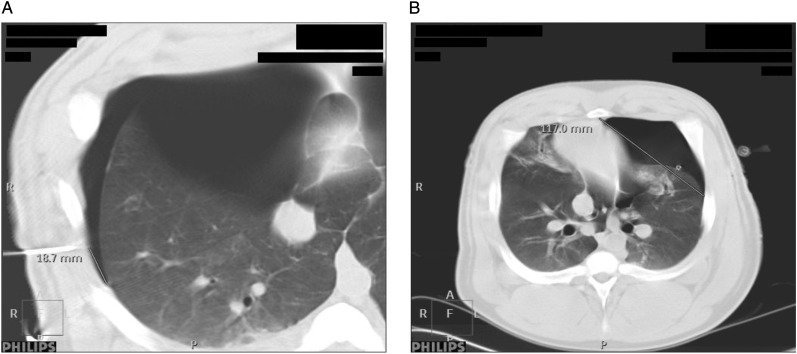
Figure 4.
A, Difference in lung point localization was determined by comparing the ultrasound-placed needles with the extension of the PTX on the CT scans. B, The sternum-lung point (S-LP) distance. The lung is increasingly separated from the chest wall as the PTX expands, leading to lateralization of the lung point and increased S-LP distance. See Figure 2 legend for expansion of other abbreviations.

