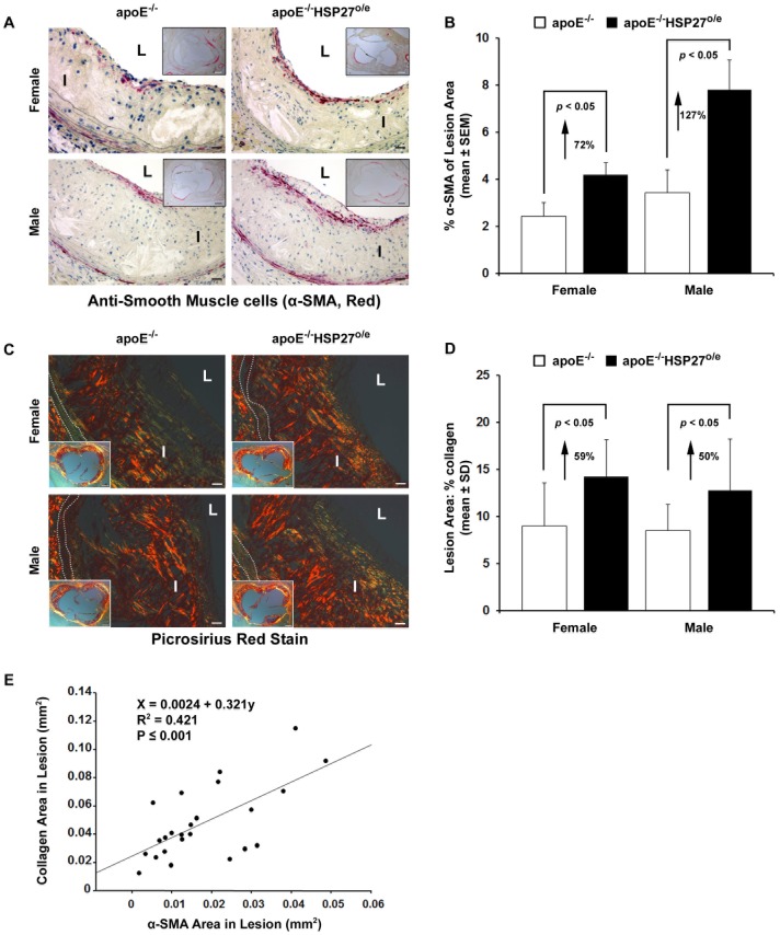
Figure 5. Increased intimal SMC and collagen content with HSP27 over-expression.
(A) SMC (anti-α-SMA red immunolabeling reaction product) localized mainly in sub-endothelial layers along the luminal surface of aortic sinus cross-sections. (B) Quantification of SMC content in aortic sinus cross-sections. (C) Intimal collagen content demonstrated by polarized microscopy. Bright yellow or orange birefrigence of collagen, due to co-aligned molecules of Type I collagen. (D) Quantification of intimal collagen content. (E) Correlation between collagen and SMC content in lesions. Scale bar in (A) and (C) = 20 µm and 200 µm (insert photo), L = Lumen, I = Intima, dotted lines delineates the media.

