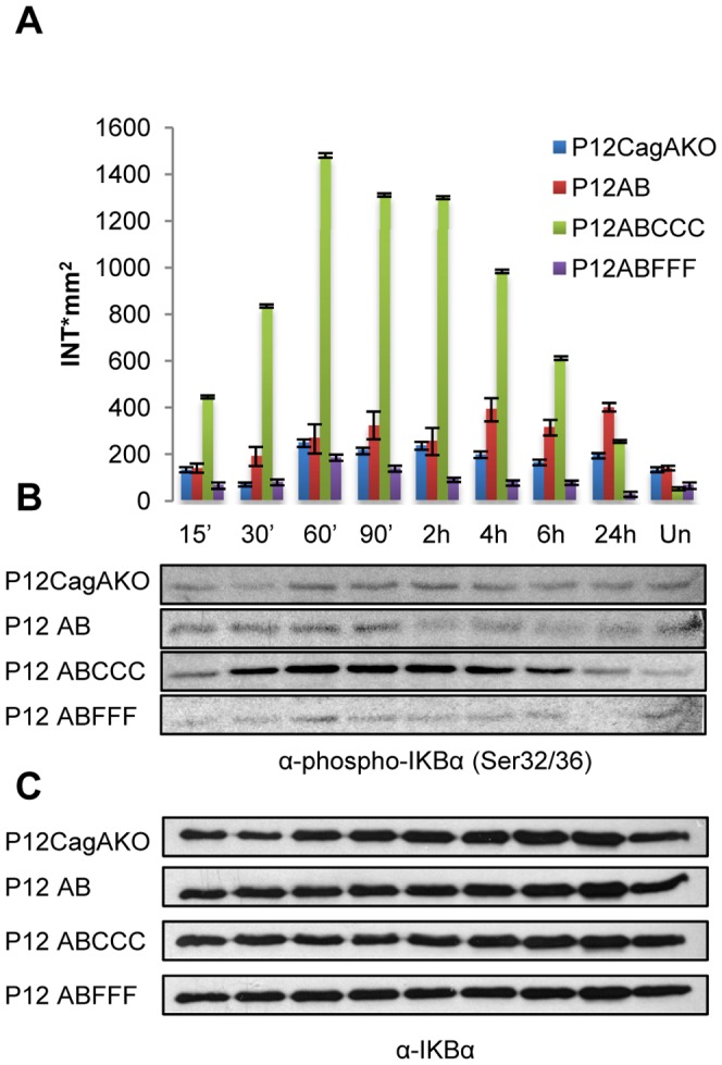
Figure 3. NF-kB activation in AGS cells following H. pylori infection.

IkBα phosphorylation at Ser32/36 in AGS cells infected with H. pylori mutants, expressing CagA with phosphorylation-functional (EPIYA-C) or -defective (EPIFA-C) motifs. (A) Quantification of IkBα phosphorylation determined by band densitometry utilizing Quantity One software package. (B) Phosphorylation of IkBα and (C) Expression of total IkBα. Un: uninfected cells.
