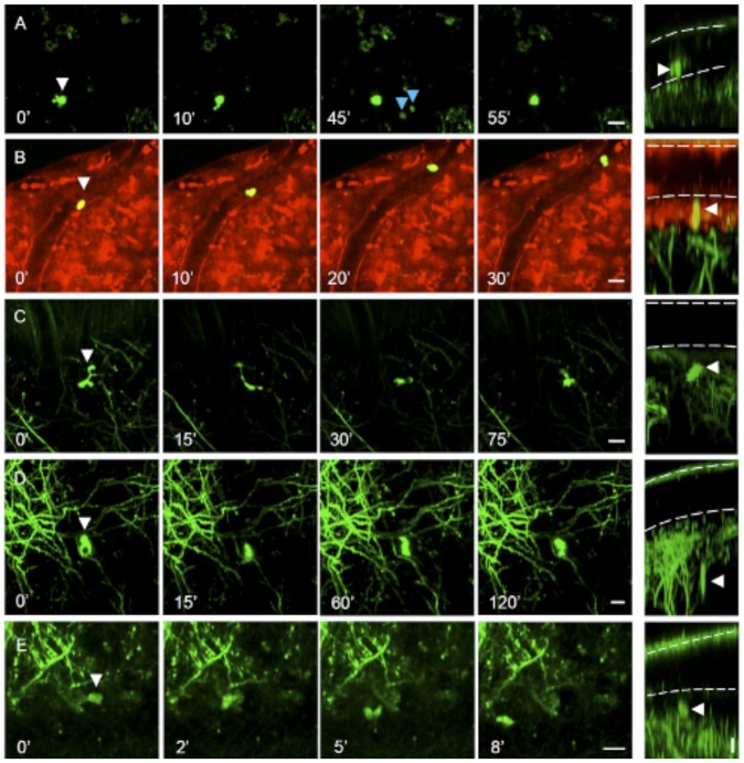
Figure 1. In vivo observation of motile GFP-labeled cells in the cortex of thy1GFP-M mice.
(A) GFP+ cell (white arrowhead) above the pial surface rapidly changing its morphology. Time-lapse sequence of maximum intensity projections of a set of optical sections acquired at 2 µm z-step. The right column shows the depth of the cell through a digital rotation of the corresponding images on the left. The white dotted lines indicate the dura and pia mater.The frame acquired at 45′ shows fluorescent cells (blue arrowheads) passing above the pia through the CSF. (B) GFP+ cell rolling inside a blood vessel on the pial surface. The blood vessel walls (shown in red) were labeled by the intravital dye SR101. Fluorescent cells showing motility at the pial surface (C) and deep in the brain parenchyma (D–E). E) GFP+ cells showing translation across the field of view. Scale bars 10 µm.

