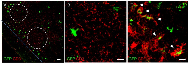
Figure 5. GFP-DCs in cervical lymph nodes of a thy1GFP-M mouse.

(A) Confocal analysis of cryosectioned cervical lymph node shows the presence of numerous GFP-tagged cells (green). They surround the B cell follicles (dashed lines) and extend in the T cell zone (on the upper right of the figure), while they rarely occur in the subcapsular zone. CD3+ cells, visualized by immunohistochemistry, are here shown in red. (B) High magnification of CD3+ cells (red) and GFP+ cells (green) in the T cell zone. (C) Immunopositivity of GFP-tagged cells (green) to CD11c (red; white arrowheads). Note that the GFP is expressed in the cytoplasm of GFP+DCs.
