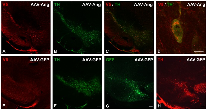
Figure 3. Angiogenin is expressed in TH-positive neurons in the substantia nigra after viral injection.
A. An AAV2-Ang injected mouse was sacrificed eight weeks after injection and stained with a primary antibody against the V5 tag fused to angiogenin and a CY3-conjugated secondary antibody, demonstrating overexpression of angiogenin in the substantia nigra. Scale bar = 100 µm. B. Same section of tissue stained with a primary antibody against tyrosine hydroxylase (TH) and an Alexa488-conjugated secondary antibody to identify dopaminergic nigral neurons. Scale bar = 100 µm. C. A merge of V5-angiogenin staining and TH staining demonstrating colocalization in dopaminergic cells in the substantia nigra. Scale bar = 100 µm. D. Higher magnification image demonstrating V5-angiogenin localized perinuclearly and accumulated in vesicular-like puncta within the cell. Scale bar = 10 µm. E. Control immunostaining of the substantia nigra from an AAV2-GFP injected mouse. No V5-angiogenin staining was observed. Scale bar = 100 µm. F. Same section stained for TH to identify dopaminergic neurons in the substantia nigra of an AAV2-GFP-injected mouse. Scale bar = 100 µm. G. GFP immunostaining of the substantia nigra from another section from the same AAV-GFP injected mouse, demonstrating GFP expression in the substantia nigra. H. Same section stained for TH to identify dopaminergic neurons in the substantia nigra of an AAV-GFP injected mouse. Scale bar = 100 µm.

