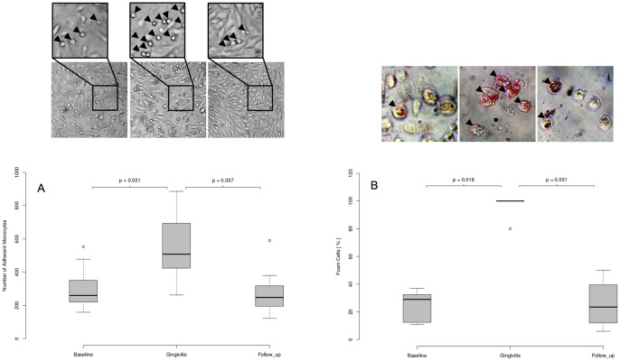
Figure 5. Monocyte adhesion assays and foam cell formation at baseline, gingivitis and follow-up.
Blood monocytes were isolated from the blood of the volunteers at baseline (day 0), gingivitis (day 21) and follow-up (day 42) and subjected to ex vivo activation assays. (A) Number of adherent monocytes on cultured endothelial cells (HUVECs). Arrowheads in the enlarged picture detail indicate adherent monocytes. (B) Percentage foam cell formation after stimulation with oxLDL (10 µg/ml). Arrowheads indicate oil red O-positive foam cells. Representative pictures are shown. P-values were calculated using two-sided Wilcoxon signed-rank tests for paired samples.

