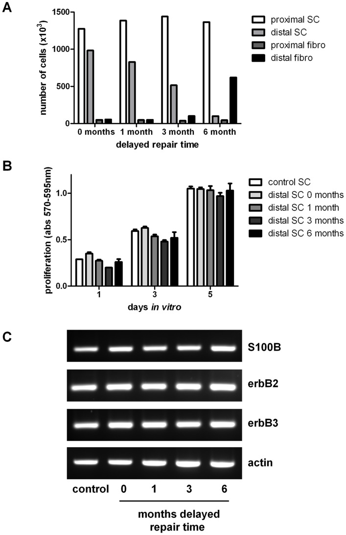
Figure 5. Characterisation of Schwann cells isolated from nerve segments.
(A) The number of Schwann cells from proximal and distal nerve stumps of animals undergoing sciatic nerve injury followed by either immediate or delayed nerve repair (at the time points indicated) with a 10 mm graft were counted 7 days following enzyme digestion of the nerve. Fibroblast counts were also made following treatment of the samples with magnetic anti-fibroblast beads. (B) Distal nerve segment Schwann cells from (A) were trypsinised and replated and proliferation rates (in the presence of glial growth factors) compared with control normal cultures of Schwann cells (no experimental injury or repair). (C) Qualitative RT-PCR analysis of Schwann cell marker S100 and the glial growth factor receptors erbB2 and erbB3 expression levels in cultured cells isolated from control nerve (no experimental injury or repair) and the distal nerve segments of animals undergoing immediate (0 months) or delayed nerve repair (1, 3, 6 months). Actin was used as a house-keeping gene.

