
Figure 2. Blockade of sEPCR binding to monocytes by anti-EPCR and anti-Mac-1 in flow cytometry.
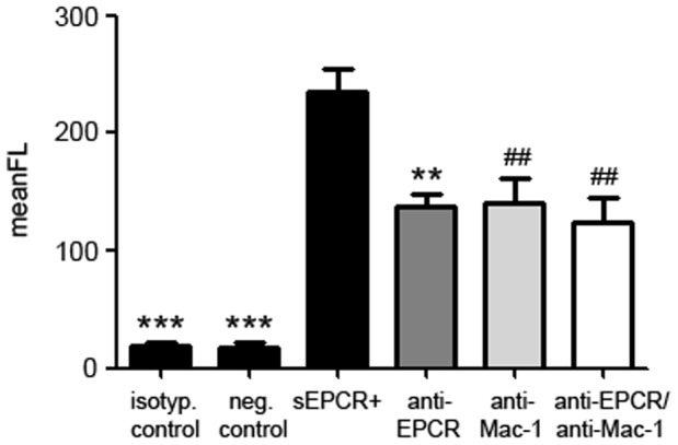
Binding of soluble, recombinant EPCR to monocytes (black bar second from left) can be blocked by anti-EPCR (dark grey bar), by anti-Mac-1 (light grey bar) and by both antibodies (white bar) to an equal extend. Monocytes without addition of sEPCR and isotype IgG served as negative controls (black bars on the left and second from left). (*** p<0.0001; ** p<0.001; ## p<0.005 vs. sEPCR+).