
Figure 4. Blockade of monocyte binding to endothelial cells by anti-EPCR and anti-Mac-1 under flow conditions.
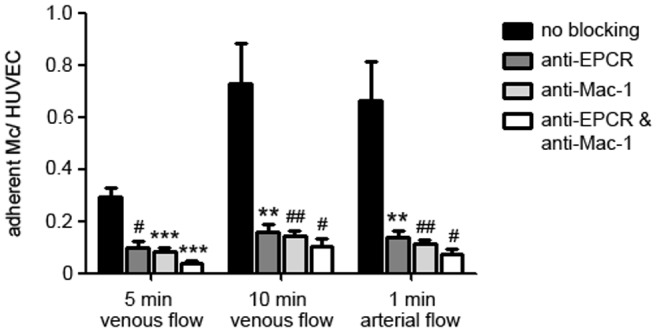
Monocyte binding to HUVECs in a dynamic adhesion assay after 5 min (left) and 10 min venous flow (middle), and after 1 min arterial flow (right). When EPCR was blocked on HUVECs (dark grey bars), Mac-1 on monocytes (light grey bars), or both (white bars) monocyte binding could be significantly diminished compared to native cells without antibody treatment (black bars). (*** p<0.0001; ** p<0.001; ## p<0.005; # p<0.05 vs. no blocking).