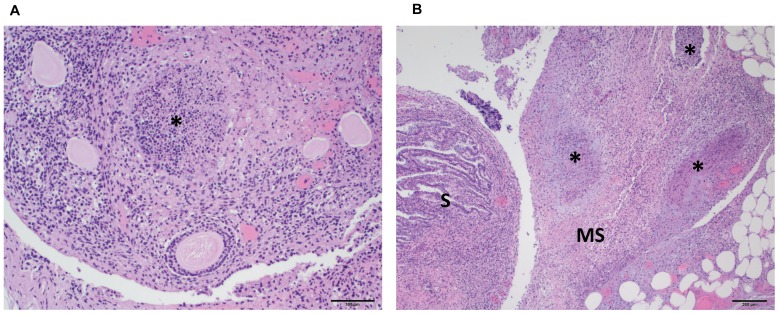
Figure 9. H&E stained reproductive tissues collected from a prairie dog that died due to MPXV infection (PD8021).
Figure 9a shows a low magnification photomicrograph of the ovary with a necrotic focus (*) and associated inflammatory infiltrate (100× original magnification, scale bar = 100 micrometer). Figure 9b shows a low magnification photomicrograph of the salphynx (S) and adjacent mesosalphynx (MS) (40× original magnification, scale bar = 200 micrometer). There is extensive necrosis and pockets of inflammation (*) in the mesosalphynx.

