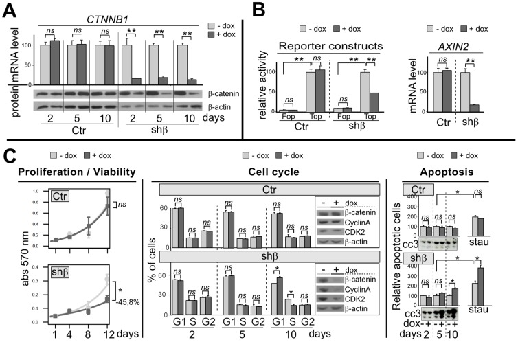
Figure 1. CTNNB1 silencing alters the Wnt/β-catenin signaling pathway, proliferation, cell cycle and apoptosis.
A, Histogram and Western blot panels represent CTNNB1 (β-catenin) mRNA and protein accumulation, in Ctr and shβ clones after 2, 5 or 10 days after addition of doxycyclin (dox) in the culture medium (0.2 µg/ml). B-left, cells were transiently co-transfected with an artificial Wnt/-β-catenin pathway reporter construc (Top) or his control mutated (Fop). After 24 h, cells were treated by vehicle or dox (0.2 µg/ml) for 24 h and luciferase activity was measured. -right, Histogram represent AXIN2 mRNA accumulation after 2 days of dox treatment. C-left, Cell survival curve of cells, as assessed by the MTT assay without or with dox (0.2 µg/ml) for 1, 4, 8 or 12 days. -center, The distribution of cells in the various phases of the cell cycle was analysed by flow cytometric analysis of propidium iodide staining after vehicle or dox treatment for 2, 5 and 10 days. Western blots show β-catenin, CyclinA and CDK2 protein levels at 10 day. -right, Histograms represent apoptotic cells measured by flow cytometric annexin V incorporation, after vehicle or dox treatment for 2, 5 and 10 days, without or with staurosporin co-treatment for last 6 h (0.5 µg/ml). Western blots show the cleaved caspase 3 (cc3) in same condition.

