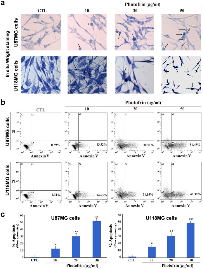
Figure 2. Determination of induction of morphological and biochemical features of apoptosis in human glioblastoma cells following photofrin based PDT.
Treatments: control (CTL), 10, 20, and 50 µg/ml photofrin incubation for 4 h followed by irradiation with 670 nm light dose of 1 J/cm2. (a) In situ Wright staining to examine morphological features of apoptosis. (b) Annexin V-FITC/PI double staining and flow cytometric analysis of apoptotic populations after the treatments. Photofrin based PDT induced significant population of cells in A4 area, indicating induction of a biochemical feature of apoptotic death. (c) Determination of percentages of apoptosis based on biochemical feature revealed by Annexin V-FITC staining. Significant difference between CTL and a treatment was indicated by *P<0.05 or **P<0.01.

