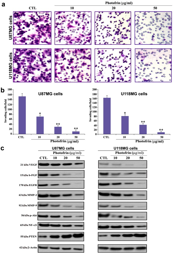
Figure 3. Changes in capability of cell invasion and alterations in expression of angiogenic, invasive, and survival factors in glioblastoma cells after photofrin based PDT.
Treatments: control (CTL), 10, 20, and 50 µg/ml photofrin incubation for 4 h followed by irradiation with 670 nm light dose of 1 J/cm2. (a) Representative Matrigel invasion assay (48 h) using U87MG and U118MG cells. A significant reduction in the number of invaded cells indicated the decrease in invasive capability of the cells after dose-dependent photofrin based PDT. (b) Quantitative evaluation of Matrigel invasion. Data indicate mean ± SEM of 10 randomly selected microscopic fields from 3 independent wells. Significant difference between CTL and a treatment was indicated by *P<0.05 or **P<0.01. (c) Western blotting using the primary IgG antibodies against VEGF, b-FGF, EGFR, MMP-2, MMP-9, p-Akt, NF-κB, and PTEN, and β-actin (loading control).

