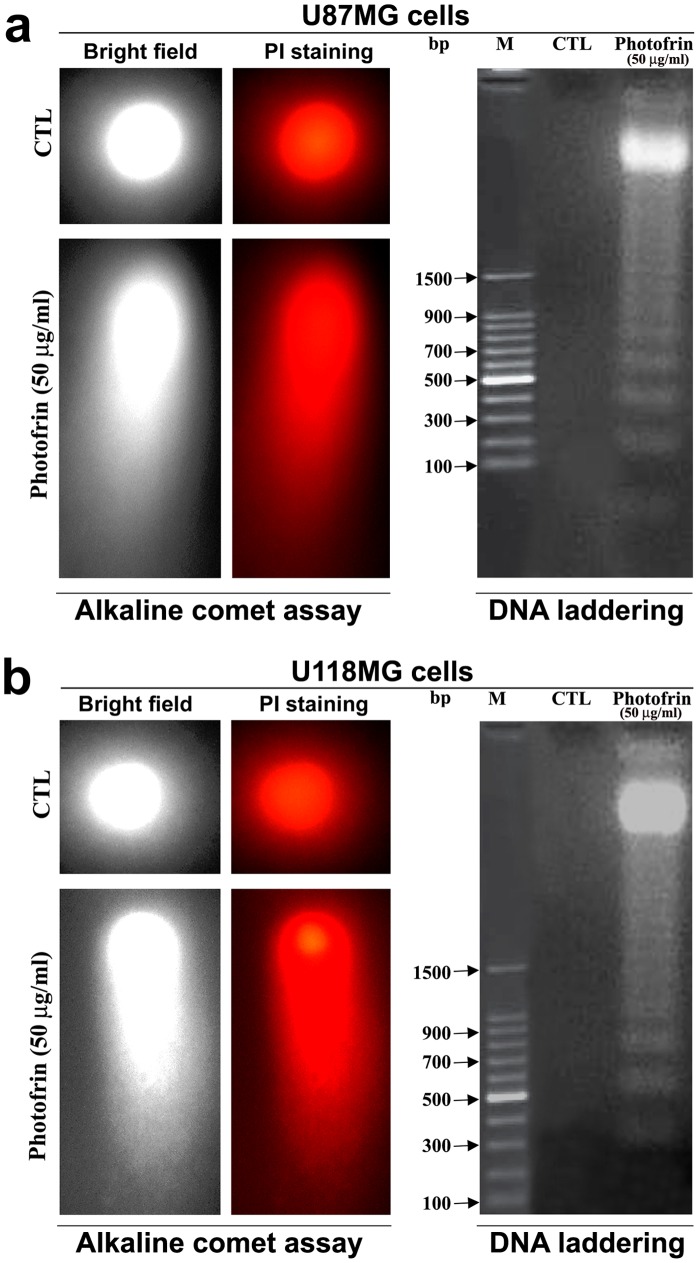
Figure 5. Alkaline comet assay and agarose gel electrophoresis to examine DNA fragmentation patterns in U87MG and U118MG cells after photofrin based PDT.
(a) Photomicrographs showing the DNA fragmentation patterns in U87MG cells. Two U87MG cells, one control (CTL) and another cell with highly damaged DNA due to photofrin based PDT, are shown in alkaline comet assay. Also, cells were treated with 50 µg/ml photofrin and irradiated with 670 nm light (1 J/cm2) and incubated for 3 h before isolation of total genomic DNA for DNA laddering assay. The CTL showed intact DNA whereas DNA ladder appeared due to photofrin based PDT. (b) Photomicrographs showing the DNA fragmentation patterns in U118MG cells. Two U118MG cells, one CTL and another cell with highly damaged DNA due to photofrin based PDT, are shown in alkaline comet assay. Also, cells were treated with 50 µg/ml photofrin and irradiated with 670 nm light (1 J/cm2) and incubated for 3 h before isolation of total genomic DNA for DNA laddering assay. The CTL showed intact DNA whereas DNA ladder appeared due to photofrin based PDT.

