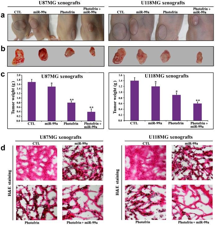
Figure 8. Regression of U87MG and U118MG tumors in nude mice and histopathological changes in tumor sections.
(a) Nude mice with U87MG and U118MG xenografts, (b) representative tumors removed surgically, (c) determination of tumor weight, and (d) evaluation of histopathological changes after the treatments. Mice with xenografts were treated for 11 days. Treatments: control (CTL) did not receive any treatment but tumor bearing mice (10th day after tumor implantation) were injected with photofrin (10 mg/kg) by tail vein, and 24 h later, 670 nm light was delivered to the tumor with fluencies of 100 J/cm2 at a fluency rate of 50 mW/cm2. We used 100 J/cm2 to expose most of the tumor cells to radiation (32). Then, the mixture of miR-99a mimic (50 µg) and 0.05% atelocollagen in 200 µl was injected (via tail vein) into each mouse on 14th, 17th, and 20th days. All animals were sacrificed on 21st day. We used 6 animals per group. Significant difference between CTL group and a treatment group was indicated by *P<0.05 or **P<0.01.

