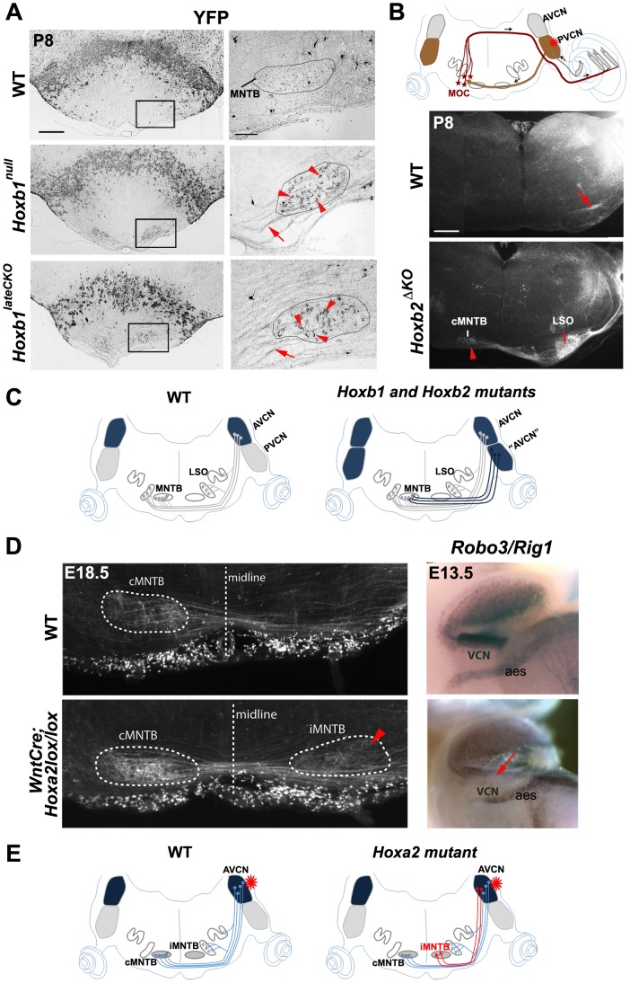
Figure 5. Abnormal cochlear connectivity in Hoxb1, Hoxb2 and Hoxa2 mutants.
(A) Oblique P8 coronal sections show abnormal presence of YFP+ fibers projecting to the medial nuclei of the trapezoid body (MNTB) in Hoxb1null and Hoxb1lateCKO mutants. Right panels: enlarged views of the boxed areas to the left depicting abnormal YFP+ terminals (arrowheads) of labeled crossed fibers (arrows) surrounding cells of the MNTB (limited by solid line). (B) Schematic representation showing the position of the injected dextran at the level of the PVCN (asterisk). Normally, PVCN interneurons innervate contralateral MOC neurons, as indicated in the control coronal section (arrow). In Hoxb2ΔKO mutants, projections originating from the PVCN target now AVCN-specific targets, such as the contralateral MNTB (cMNTB) (arrowhead) and the lateral superior olivary (LSO) nuclei. (C) Schematics summarizing the normal connectivity pattern of cochlear AVCN neurons towards the nuclei of the superior olivary complex (SOC) complex, and the abnormal presence of YFP+ neurons behaving like AVCN neurons when Hoxb1 or Hoxb2 are inactivated. (D) A subpopulation of AVCN-labeled axons project abnormally to the ipsilateral MNTB (see arrowhead) when Hoxa2 is conditionally inactivated in the Wnt1+ (rhombic lip) domain. Expression of the Slit receptor Rig1 (or Robo3) is decreased in the VCN, indicating that absence of Rig1 affects midline crossing of AVCN projections. (E) Schematics summarizing the abnormal axonal projections of AVCN neurons in Wnt::Cre;Hoxa2lox/lox mice after dextran injection in the AVCN. AVCN, anterior ventral cochlear nucleus; PVCN, posterior ventral cochlear nucleus; PVCN IN, PVCN interneurons; MOC, medial olivocochlear neurons; aes, anterior extramural stream. Scale bars: 400 µm (A left panels, B), 50 µm (A right panels).

