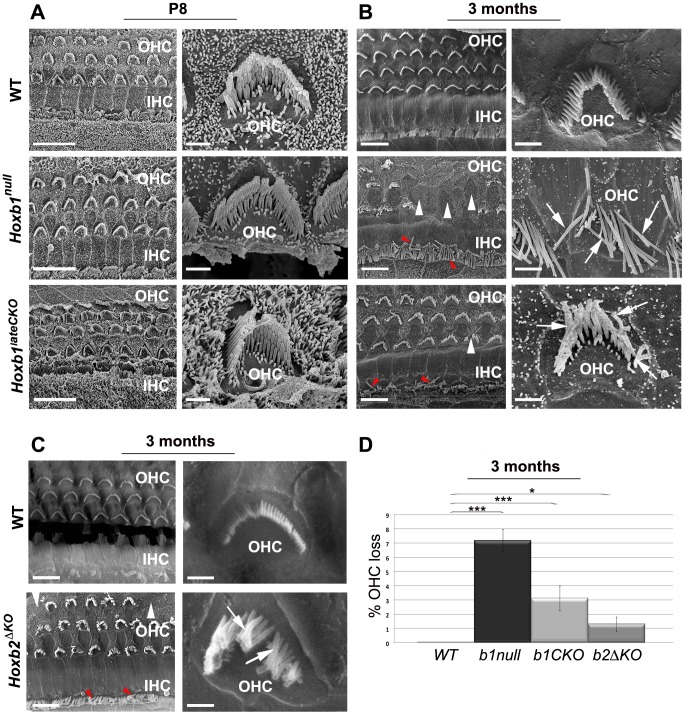
Figure 7. Late degeneration of OHCs in the apical turn of Hoxb1 and Hoxb2 mutant cochleae.
(A) Scanning electron microscopy (SEM) views of the cochlea at P8: an overview of the apical turns of WT, Hoxb1null and Hoxb1lateCKO cochleae showing three orderly arrayed rows of outer hair cells (OHCs) and one row of inner hair cells (IHCs). Representative high magnification images illustrate stereocilia of hair bundles of single OHCs arranged according to their different lengths. Shape and organization of OHCs in apical regions are normal at this stage in both mutants. (B) SEM views of 3-month-old WT, Hoxb1null and Hoxb1lateCKO cochleae and representative higher magnification images of OHCs. In Hoxb1null and Hoxb1lateCKO cochleae, OHCs have lost their regular organization and fail to develop in some areas (white arrowheads). Moreover, in Hoxb1null cochleae most stereocilia have completely lost their typical V-shaped morphology and their characteristic differences in lengths (arrows). OHCs are less severely affected in Hoxb1lateCKO cochleae. IHC cilia appeared weakly disarranged (red arrowheads). (C) SEM views of 3-months-old WT and Hoxb2ΔKO mutant cochleae and higher magnifications of representative OHCs. Note that, similarly to Hoxb1lateCKO cochleae, Hoxb2 mutants have occasional missing OHCs (white arrowheads), disarranged IHC cilia (red arrowheads) and disorganized OHC stereocilia (arrows). (D) Histogram quantifying the percentage of OHC loss in controls, Hoxb1null, Hoxb1lateCKO and Hoxb2ΔKO cochleae. While controls (n = 8) showed no OHC loss, in Hoxb1null mutants (n = 6) 7.2±0.8% of OHCs were absent, whereas 3.1±0.9% and 1.3±0.5% were lost in Hoxb1lateCKO (n = 6) and Hoxb2ΔKO (n = 3) cochleae, respectively. Inter-genotype ANOVA p<0.001; Hoxb1null versus WT: p<0.001; Hoxb1lateCKO versus WT: p<0.001; Hoxb2ΔKO versus WT: p = 0.02. Scale bars, 10 µm (A, B, C, left panels), 1 µm (A, B, C, right panels). See also Figure S8.

