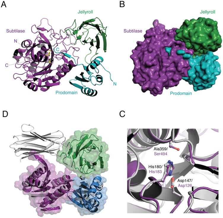
Figure 3. Overall structure of CspB perfringens.
(a) Ribbon representation showing subtilase domain in purple, jellyroll domain in green, and prodomain in teal extending into the active site. Catalytic residues are shown as stick models with yellow carbons. (b) Close-up view of catalytic site. An overlay of CspB (purple) and Tk-SP (grey). The three catalytic residues are shown. Tk-SP and CspB catalytic residues are labeled in black and purple, respectively. (c) Space-filling model of CspB with same orientation and color scheme as (a). (d) Overlay of CspB (colors, same as (a)) and Tk-SP (shown in grey), showing similar overall structures with the exception of the position of the jellyroll domain. The jellyroll domains of CspB and Tk-SP are shown in green and grey, respectively. Note that only the regions with conserved secondary structure in the prodomain and subtilase domain are shown.

