
Fig. 4. Gel mobility shift analysis of WT and mutant CsrA protein.
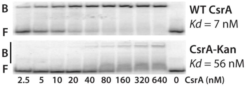
5’ end-labeled RNA was incubated with the concentration of WT or mutant CsrA shown at the bottom of each lane. Positions of bound (B) and free (F) RNA are shown.
Official websites use .gov
A
.gov website belongs to an official
government organization in the United States.
Secure .gov websites use HTTPS
A lock (
) or https:// means you've safely
connected to the .gov website. Share sensitive
information only on official, secure websites.
5’ end-labeled RNA was incubated with the concentration of WT or mutant CsrA shown at the bottom of each lane. Positions of bound (B) and free (F) RNA are shown.