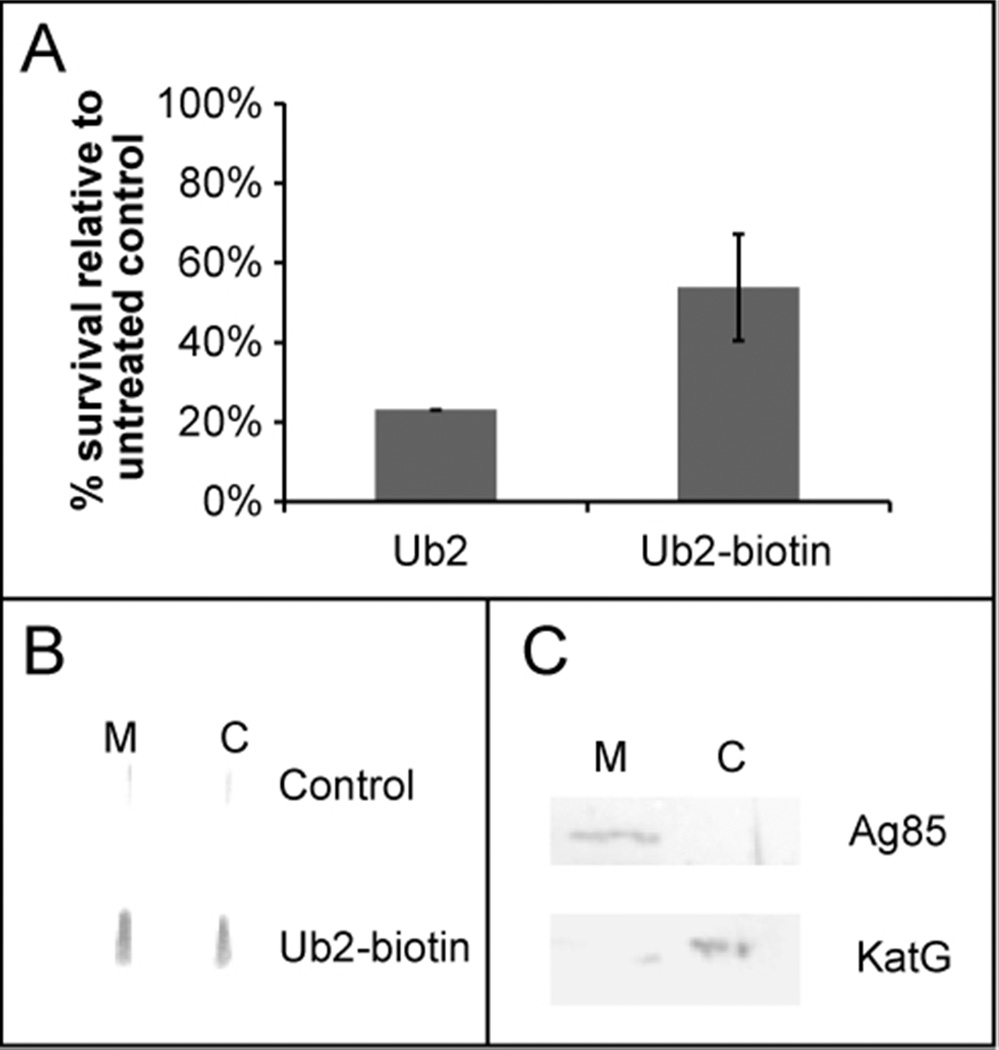
Figure 3. Subcellular localization of Ub2-biotin.
A) M. smegmatis mc2155 was sub-cultured to 5×10⁵ cfu/mL in 7H9 medium containing 50 µM Ub2 or Ub2-biotin. Following overnight treatment, the number of viable bacteria was determined by plating serial dilutions, and the average of three replicates is shown. B) M. smegmatis mc2155 was treated with 50 µM Ub2-biotin or buffer control. Bacteria were harvested, membrane (M) and cytoplasmic (C) fractions obtained and applied to nitrocellulose using a slot blotter. Western analysis was performed with monoclonal antibody against biotin. C) To assess the purity of the subcellular fractionation protocol, membrane and cytoplasmic fractions were resolved on a 12% SDS-PAGE gel and Western analysis performed using antibodies against the Ag85 membrane protein and the cytoplasmic protein KatG.

