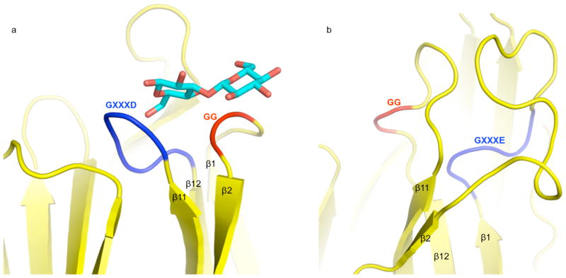
Figure 5. Glycan-binding motifs.
a. The β1β2 loop of BanLec containing the GG motif (red) and the β11β12 loop containing the GXXD motif (blue) in ribbon representation. The structure of bound laminaribiose is shown in bonds representation with carbons in cyan and oxygens in red.
b. The β1β2 loop of Cry5B domain II containing the GXXE motif (blue), and the β11β12 loop containing the GG motif (red) in ribbon representation.

