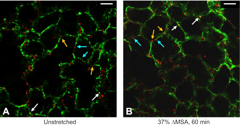
Fig. 8.
Representative live/dead stain images for day 2–3 rat PCLSs that remained unstretched (A) or stretched for 60 min at 37% ΔMSA (B). In these preparations, we added the dead cell probe ethidium homodimer-1 immediately before stretch, then stretched the PCLSs, and 1 h after stretch we added the live cell probe calcein AM to the wells. White arrows point to double stained cells. Light blue and orange arrows point to distinctive locations of alveolar epithelial cells, which may be indicative of type I and type II cells, respectively. Bar = 50 μm.

