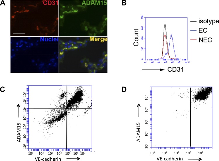
Fig. 1.
The endothelium is a predominant source of ADAM15 in lung. A: ADAM15 colocalizes with CD31 in lung tissue. Immunofluorescence microscopy demonstrates colocalization of CD31 and ADAM15. Lung sections were labeled with CD31 and ADAM15 antibody, followed by rhodamine and FITC-conjugated second antibody, respectively. Nuclei are counterstained with Hoechst 33342 (blue). Images are captured with Zeiss Axio Observer 200M inverted microscope equipped with Zeiss AxioVision software (scale bar = 25 μm). B: FACS analysis: primary lung endothelial cells (EC) or non-EC (NEC) isolated from wild-type (WT) mice were labeled with an anti-CD31 antibody or IgG isotype control. C: FACS analysis of ADAM15 demonstrated coexpression with VE-cadherin positive endothelial cells vs. NEC. D: after CD31 selection, EC cells were cultured to confluence and assayed for VE-cadherin and ADAM15 expression as shown.

