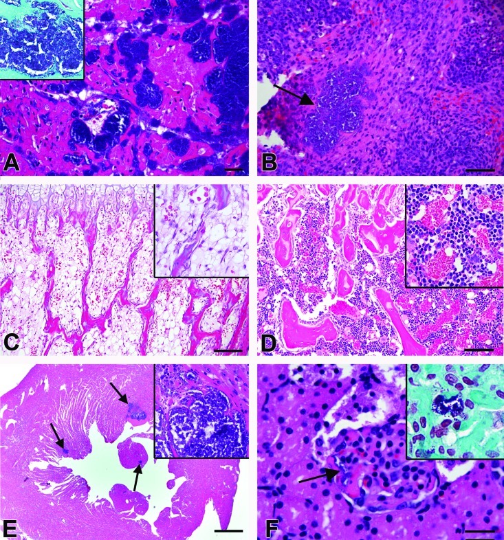
Figure 1.
Histopathology. (A) Heart case 1. Multifocal myofibrillar degeneration and necrosis accompanying large clusters of coccoid bacteria without accompanying inflammation. Hematoxylin and eosin stain; bar, 20 μm. Inset, Gram stain of heart showing gram-positive cocci within the ventricular myocardium. (B) Spleen, case 1. Focal aggregate of coccoid bacteria (arrow) without accompanying inflammation. Hematoxylin and eosin stain; bar, 50 μm. (C) Bone marrow, case 1. Marrow is hypocellular; bar, 100 μm. Inset, Higher magnification (600×) view of the bone marrow. Hematoxylin and eosin stain. (D) Normal age-matched rat bone marrow; case 2. Bar, 100 μm. Inset, Higher magnification (200×) view of the bone marrow. Hematoxylin and eosin stain. (E) Heart; case Number 9. Multifocal botryoid clusters of bacterial cocci (arrows) surrounded by degenerate and necrotic cardiac myocytes. Inflammatory cells were not associated with the cardiac lesions. Bar, 500 μm Inset, Higher magnification (600×) view of the heart. Multifocal clusters of bacteria are present within the ventricular myocardium. Hematoxylin and eosin stain. (F) Glomerulus, case 15. Clusters of coccoid bacteria (arrow) are present within the capillaries. Hematoxylin and eosin stain; bar, 20 μm. Inset, Gram stain of the kidney showing gram-positive cocci within the glomerular capillaries.

