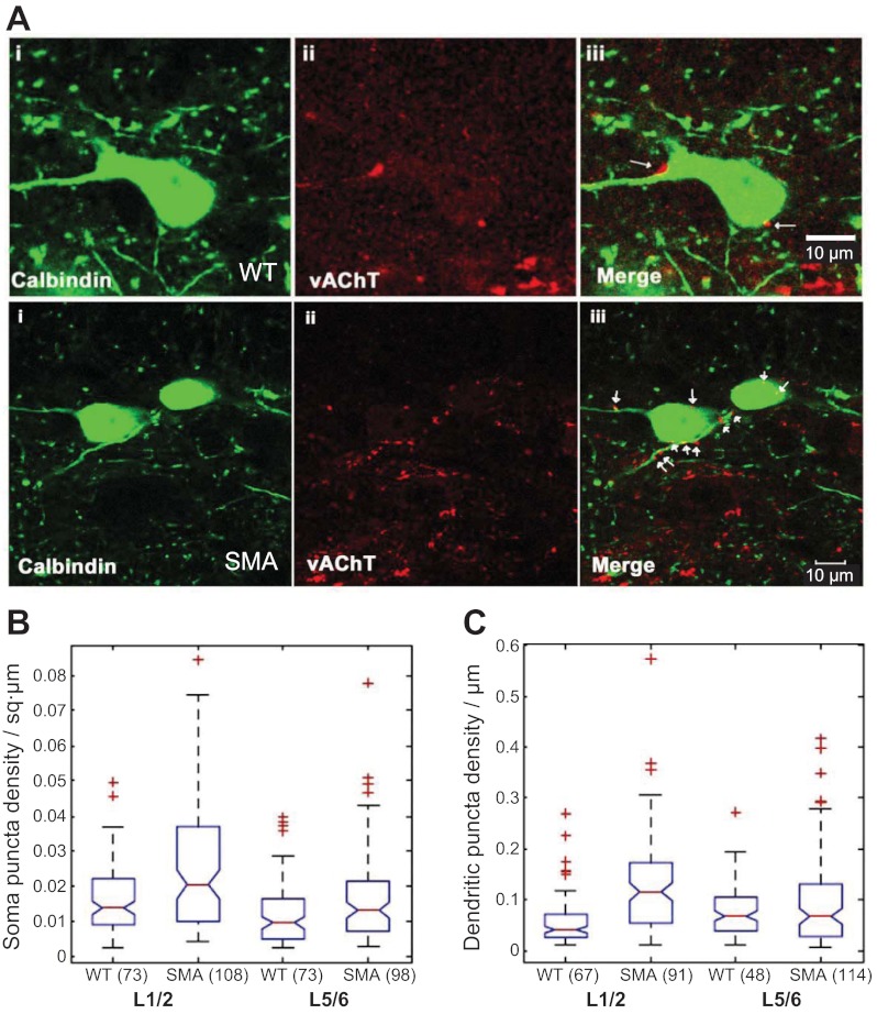
Fig. 4.
Vesicular acetylcholine transporter (VAChT+) terminals are not reduced on calbindin+ putative Renshaw cells in SMAΔ7 mice at P12–13. A, top: calbindin+ cell (i), VAChT+ terminals (ii), and a merged image (iii) at the L1/L2 segmental level of a wild-type mouse at P12–13. A, bottom: the same immunocytochemistry for a calbindin+ cell from an SMAΔ7 mouse at P12–13. Cholinergic synapses (arrows) are identified by the apposition of VAChT immunoreactive boutons to calbindin+ dendrites and somata in the merged images (iii). Images are generated from a single optical section (0.54 μm). B: box plots showing medians and IQRs of somatic VAChT puncta in wild-type and SMAΔ7 mice in the L1–L2 and L5–L6 regions. C: box plots showing medians and IQRs of dendritic VAChT puncta in wild-type and SMAΔ7 mice in the L1–L2 and L5–L6 regions. In both B and C, + symbols denote outliers. Numbers in parentheses indicate the number of somata (B) or dendrites (C) that were used to generate the graph.

