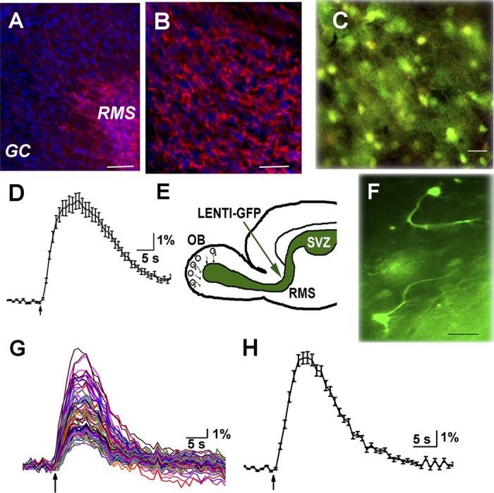
Fig. 1.

Progenitor cells in rostral migratory stream (RMS) express doublecortin and are depolarized by GABA and ACh. A: fixed frozen sections labeled with doublecortin antibody (red) showing dense labeling in RMS but not in other layers of the olfactory bulb (OB). Nuclei are stained with DAPI (blue). Scale bar, 100 μm. B: higher magnification of doublecortin labeling in RMS; scale bar, 10 μM. C: image of cells in the RMS loaded with fura 2-AM. Scale bar, 15 μm. D: calcium transients generated in a progenitor cell in the RMS upon a 10-s application of 1 mM GABA. E: cartoon of sagittal section of brain showing the site of injection. F: 2 cells in the RMS expressing GFP with processes pointing in the direction of migration. G: calcium transients in response to a 5-s puff application of 1 mM ACh and 1 μM atropine (ACh/AT). All cells in the field respond, but the response is variable among cells. H: average response from 36 cells shown in G.
