
Figure 2.
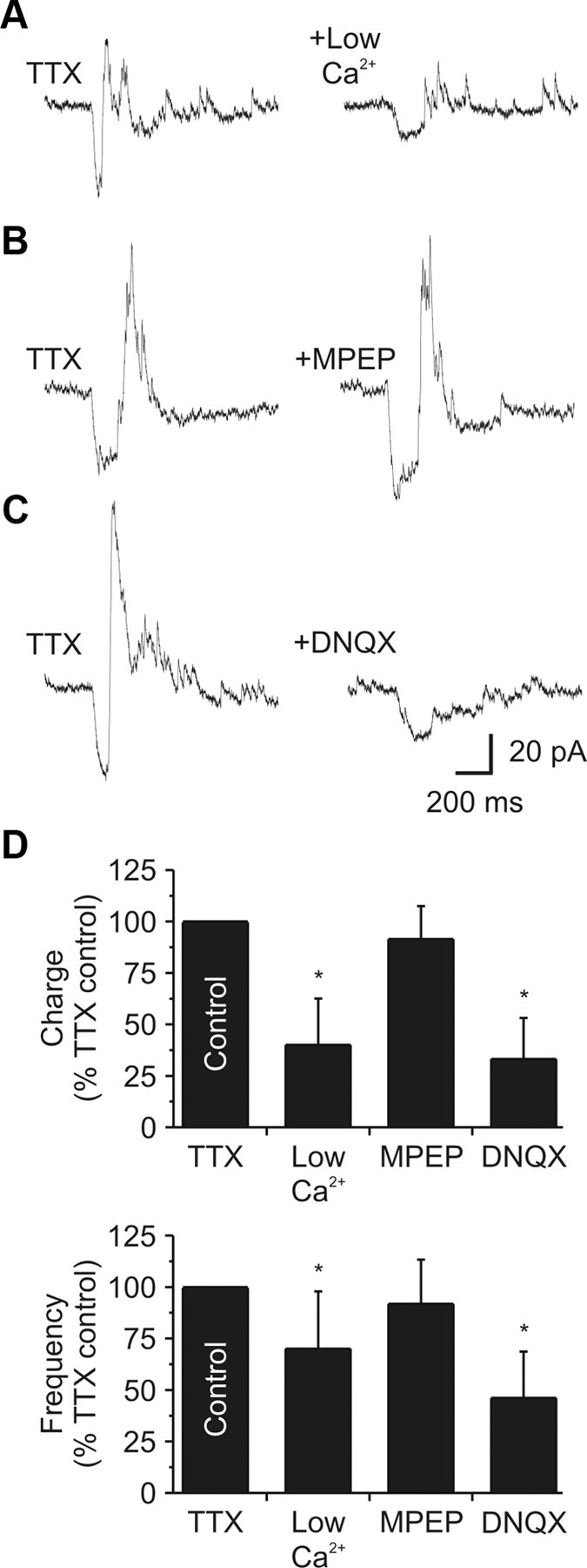
iGluRs regulate local dendrodendritic output. A, Left, In TTX (1 μm), glutamate release increased IPSC activity in a dLGN relay neuron. Right, In a low-Ca2+ (0.2 mm)/high-Mg2+ (6.0 mm) extracellular solution, the evoked IPSC activity was significantly attenuated. Responses recovered after a 10–15 min wash (data not shown). B, Left, In a different neuron (while in TTX: 1 μm), glutamate release increased IPSC activity. Right, addition of the mGluR5 antagonist MPEP (30–50 μm) did not change the evoked IPSC activity. C, Left, In a different neuron (while in TTX: 1 μm), glutamate release increased IPSC activity. Right, Addition of the AMPA receptor antagonist DNQX (20–40 μm) significantly reduced the evoked activity. D, Population data illustrating the decrease in IPSC charge and frequency by both low-Ca2+ and DNQX.