Figure 4.
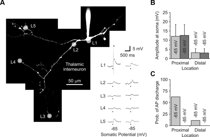
Glutamate uncaging generates local and not global GABA release from thalamic interneurons. A, Right, A 2PLSM image of a local thalamic interneuron filled with Alexa Fluor 594 (50 μm). Without TTX, glutamate uncaging (white shadows) was performed at various locations (L) while recording the voltage response at the soma. Left, Somatic voltage recordings from each location while holding the soma at a depolarized (−65 mV) and hyperpolarized (−85 mV) membrane potential. The star (*) indicates a truncated action potential, while the block dot represents when the laser pulse was delivered. A plateau-like calcium spike (Acuna-Goycolea et al., 2008) was never generated with focal glutamate stimulation. B, Population data indicating that the response amplitude was independent of holding potential and was dependent on location (Proximal: 0–44 μm; Distal: 150–263 μm). C, Population data indicate that the probability of spiking was highest when stimulating proximal locations at a depolarized membrane potential. No spiking was observed when the cell was hyperpolarized.

