Figure 6.
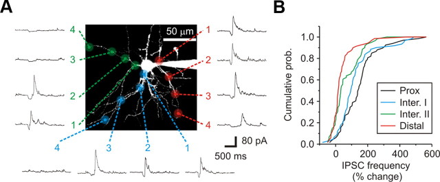
Inhibitory dendrodendritic synapses are preferentially localized to the proximal dendrites of relay neurons. A, A 2PLSM image of a dLGN relay neuron filled with Alexa Fluor 594 (50 μm). Glutamate was released at four spatially distinct locations along individual primary dendrites. As measured from the soma: (1) Proximal (Prox): 15–25 μm; (2) Intermediate I (Inter. I): 30–45 μm; (3) Intermediate II (Inter. II): 50–75 μm; (4) Distal: 80–120 μm. Shown are the corresponding responses for each of the locations tested. B, A cumulative probability graph for each dendritic location examined. The probability of observing a change in IPSC activity decreased from proximal to distal locations.

