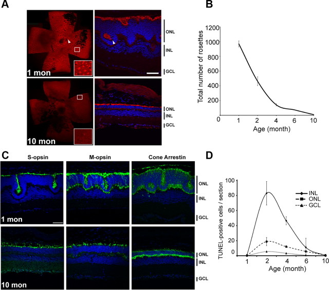
Figure 2.
Transitory cell death between 1 and 4 months in Nrl−/− retina. A, Identification of pseudorosettes with PNA on flat-mount retina and cryosections on 1- and 10-month-old Nrl−/− mice. B, Average of the number of pseudorosettes in Nrl−/− mice counted after labeling with PNA on flat-mount retina at 1, 2, 4, 6, and 10 months of ages. The maximum decrease is observed between 1 and 4 months. Error bars show ±SEM from four independent mice. C, S-opsin, M-opsin, and cone arrestin immunostaining (green) in 1- and 10-month-old Nrl−/− retina show maintenance of expression in old mice. D, By TUNEL assay, quantification of apoptotic cells per section in the outer nuclear layer (dashed line), inner nuclear layer (solid line), and ganglion cell layers (dotted line), separately in 1-, 2-, 4-, 6-, and 10-month-old Nrl−/− mice. Error bars show ±SEM from four independent mice. The data show cell death in all retinal layers between 2 and 4 months. The cell death was more pronounced in the inner nuclear layer compared with outer nuclear layer. A, C, Nuclei were visualized by DAPI staining. Scale bar, 20 μm.

