
Figure 4.
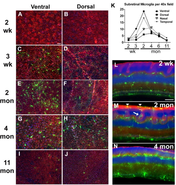
Migration of retinal microglia into the outer retina during the period of photoreceptor degeneration. Microglia migration into the subretinal space onto the RPE monolayer were imaged in sclerochoroidal flat mounts in the ventral and dorsal quadrants, respectively, at different ages: 2 weeks (A, B), 3 weeks (C, D), 2 months (E, F), 4 months (G, H), and 11 months (I, J). K, Quantification of microglia in the subretinal space demonstrates that microglia accumulation begins at 3 weeks and peaks at 2 months, with larger numbers accumulating in the ventral quadrant compared with the dorsal quadrant. RPE cell density and morphology are not significantly changed during this period. L–N, Cross-sectional views of microglia distribution in vibratome sections of the retina at different ages. At 2 months (M), microglia are closely associated with photoreceptor rosette structures (arrow) and also in the subretinal space (arrowheads). At 4 months (N), microglial density in the outer retina decreases as the photoreceptor pseudorosettes resolve and disappear. Scale bars, 50 μm.