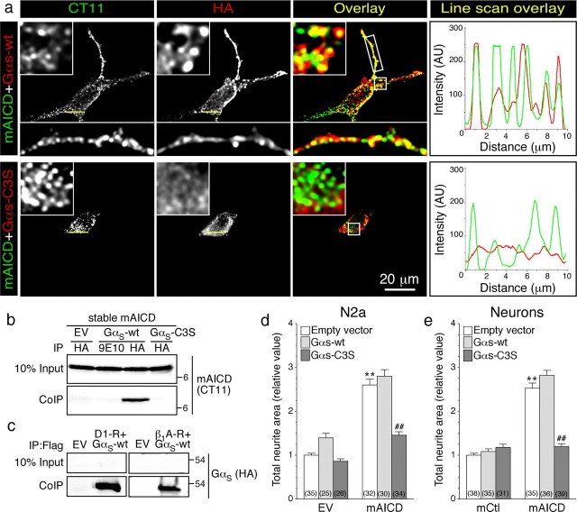
Figure 7.
Functional coupling of membrane-tethered APP intracellular domain with GαS-protein. a, Immunofluorescence localization of mAICD and HA-tagged wild-type GαS (GαS-wt) or dominant-negative GαS mutant (GαS-C3S) in N2a cells. The boxed regions are shown as enlarged insets. Line scans (right panels) show overlapping peaks of mAICD (green) and GαS-wt (red) localization along a small stretch in the cell body. b, Coimmunoprecipitation analysis of mAICD interaction with GαS was evaluated in stable N2a cells expressing mAICD following transient transfection of GαS-wt. Non-denaturing lysates were immunoprecipitated with mAb HA or mAb 9E10 (negative control) and analyzed by immunoblotting with CT11 to detect mAICD. c, As positive control, interaction between dopamine D1 receptor (D1-R) or β1 adrenergic receptor (β1A-R) and GαS-wt was analyzed by coimmunoprecipitation. d, e, Quantification of neurite outgrowth in N2a cells (d) or cortical neurons (e) 24 h following cotransfection of EV, mCtl, or mAICD with GαS-Wt or GαS-C3S. Statistical significance was examined by ANOVA Kruskal–Wallis test followed by Dunn's post hoc multiple-comparison analysis. **p < 0.001, compared with EV/EV or EV/mCtl control cells. ##p < 0.001, compared with EV within mAICD transfected group when cells are G-protein transfected. The total number of quantified cells is shown in parentheses. Error bars indicate SEM.

