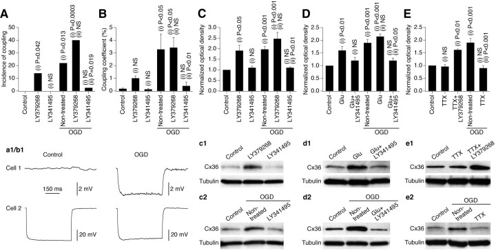
Figure 2.
Ischemic increase in neuronal gap junction coupling in vitro is regulated by glutamate via group II mGluRs and is action potential-dependent. Data from experiments in wild-type mouse mature neuronal somatosensory cortical cultures are shown. All pharmacological agents were present in the culture medium for 10 min before, during, and 10 min after OGD or sham OGD and then washed out. The analysis was done 2 h after OGD or sham OGD. A, B, Electrotonic coupling. Statistical data and representative traces (a1, b1) are shown. Presented are the incidence of electrotonic coupling and the coupling coefficient. Statistical analysis was as follows: Fisher's exact probability test (A; see Table 1 for the number of samples) and ANOVA with post hoc Tukey (B; responses from all of the tested pairs are included in the analysis). C–E, Western blots: Cx36 expression. Statistical data and representative blots (c1–e2) are shown. Optical density signals are normalized relative to tubulin and compared with the control (set at 1.0). Statistical analysis was as follows: ANOVA; n = 6–15 per group. In all graphs, statistical significance is shown relative to control (i) and nontreated plus OGD (ii). In B–E, the data are shown as mean ± SEM. Glu, Glutamate.

