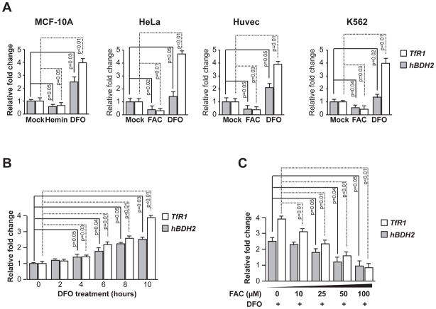
Figure 2. Iron-deficiency leads to increased hBDH2 mRNA levels.
(A) Human cell lines were treated with hemin, FAC, DFO or mock-treated for 16 hours and TfR1 and hBDH2 mRNAs were quantified by real time PCR analysis 16 hours post treatment. (B) MCF-10A cells were treated with DFO for the indicated times and hBDH2 and TfR1 mRNAs were quantified as described above. (C) Iron supplementation reverses DFO-induced increase in hBDH2 and TfR1 mRNAs. MCF-10 A cells were treated with 100 μM of DFO along with increasing amounts of FAC and relative levels of hBDH2 mRNA were quantified 10 hours post treatment as described above. For A – C, value on y-axis was set at 1.0 for the mRNA levels in untreated cells. The relative mRNA levels in each sample were normalized to actin mRNA. Results shown are the average of three independent experiments with error bars depicting SD.

