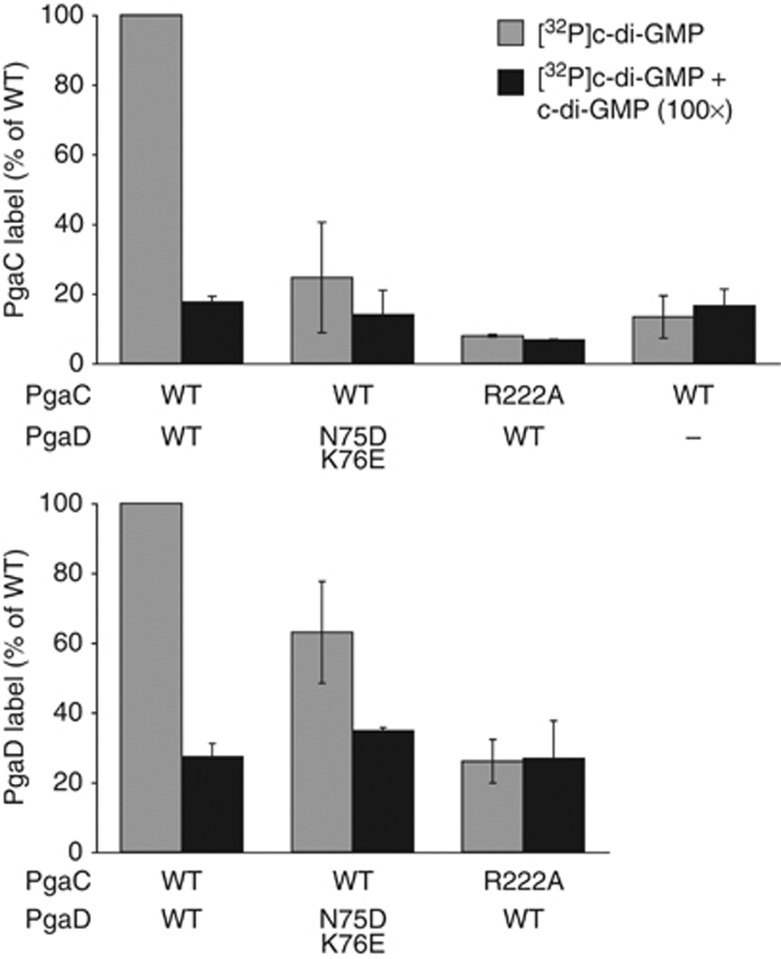
Figure 7.
C-di-GMP directly binds to both PgaC and PgaD. Membranes containing wild-type and mutant forms of PgaC–3 × Flag and/or PgaD–3 × Flag were UV crosslinked in the presence of [32P]c-di-GMP and with (black bars) or without excess c-di-GMP (grey bars). Relative PgaC (upper graph) and PgaD (lower graph) autoradiography band intensities are shown as an average of two independent experiments with standard deviations.

