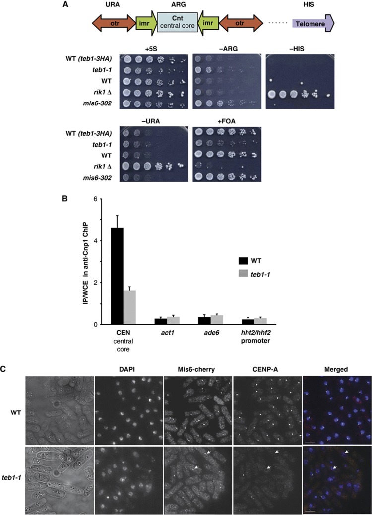
Figure 5.
Teb1 is required for normal Cnp1 loading. (A) Silencing at the centromeric central core is specifically reduced in teb1-1 cells. Strains of the indicated genotypes harbouring a ura4+ marker in the otr region, an arg3+ marker in the central core and a his3+ marker in the subtelomere were analysed by serial dilution assay. (B) Cnp1 binding to the central core decreases in teb1-1 cells compared to wt. ChIP was performed with an anti-Cnp1 antibody. The experiment was performed in triplicate and error bars correspond to the standard deviation. (C) Indirect immunofluorescence shows that while Mis6 and Cnp1 colocalize at wt centromeres, Cnp1 foci fail to appear in many teb1-1 cells; Mis6 localization is not affected (white arrowheads).

