Figure 1.
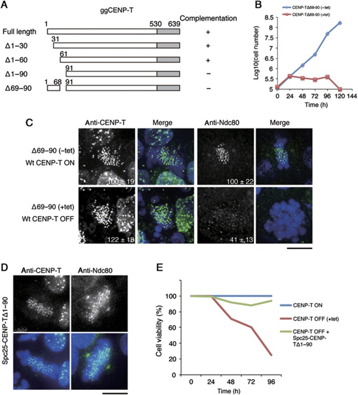
The CENP-T N-terminal region is required for kinetochore localization of the Ndc80 complex. (A) Diagram showing the chicken CENP-T sequence and the tested deletion mutants in the CENP-T N-terminal region. ‘+’ or ‘−’ indicates whether the given deletion mutant can complement growth when endogenous CENP-T is depleted. (B) Graph showing the growth curve of cells expressing CENP-TΔ69–90 in the presence (red rectangle) or absence (blue diamond) of tetracycline to repress the expression of wild-type CENP-T. (C) Immunofluorescence analysis of cells expressing CENP-T Δ69–90 after 72 h in the presence (lower panel) or absence (upper panel) of tetracycline to repress the expression of wild-type CENP-T. Cells were probed for either CENP-T or Ndc80 and the kinetochore signal intensities of each protein were measured relative to an adjacent background signal. Bar, 10 μm. (D) Immunofluorescence analysis of cells in which expression of CENP-T is replaced with a Spc25-Δ1–90-CENP-T fusion protein. Ndc80 localizes to kinetochores in these cells, unlike the CENP-T Δ69–90 mutant alone in (C). (E) Cell viability analysis for CENP-T conditional knockout cells in the presence (CENP-T OFF) or absence (CENP-T ON) of tetracycline, or expressing a Spc25-Δ1–90 CENP-T fusion protein in the presence of tetracycline (CENP-T OFF+Spc25-Δ1–90 CENP-T).

