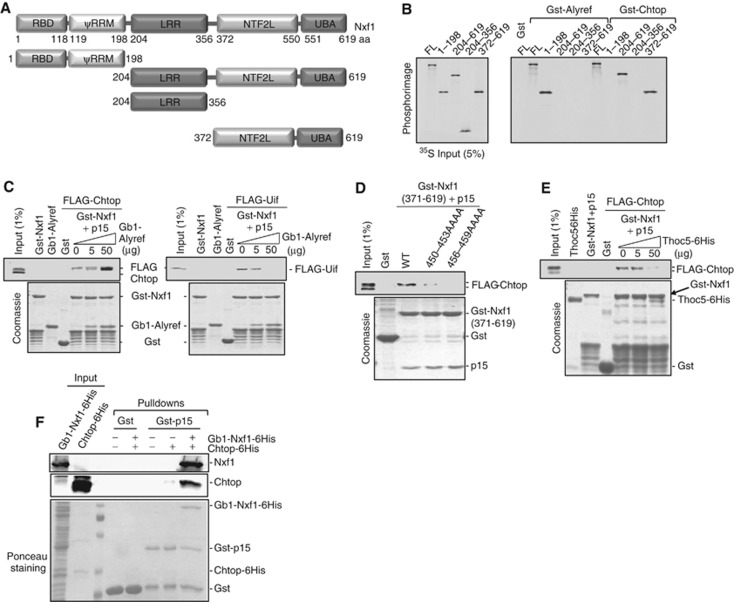
Figure 4.
Mutually exclusive binding of Thoc5 and Chtop to Nxf1. (A) Schematic representation of Nxf1 truncations used in this study. (B) Gst-Alyref and Gst-Chtop pulled down 35S-labelled Nxf1 full length and truncations. (C) Pull-down competition assay with Gst-Nxf1-p15, 293T overexpressed FLAG-Chtop or FLAG-Uif, and increasing amounts of purified Gb1-Alyref in the presence of RNase A. Proteins were detected by Coomassie staining or western blot. (D) Pull-down assays using Gst-Nxf1(aa 371–619)-p15 wild type or mutants with 293T cell extracts from cells transfected with FLAG-Chtop. Proteins were detected by Coomassie staining or western blot. (E) Pull-down competition assay with Gst-Nxf1-p15, with 293T cell extracts from cells overexpressed FLAG-Chtop and increasing amounts of purified Thoc5-6His. Proteins were detected via Coomassie staining or western blot. (F) Chtop does not bind p15 directly. Gst pulldowns with the indicated fusions were carried out in the presence of 4 μg baculovirus derived recombinant Chtop and Gb1-Nxf1 purified from E. coli as indicated. Chtop and Nxf1 were detected using monoclonal antibodies to each protein. The lower panel shows a ponceau stain of the western blot used to detect Nxf1 and Chtop. Experiments were performed in the presence of RNase A (10 μg/ml).
Source data for this figure is available on the online supplementary information page.

