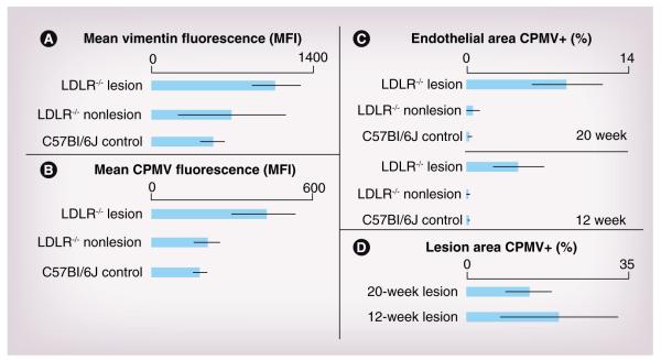
Figure 4. Quantification of vimentin and cowpea mosaic virus in lesion versus nonlesion tissue.
Analysis of vimentin and CPMV in tissue sections using ImageJ. (A) Mean fluorescence of vimentin staining in the endothelium for C57Bl/6J control mice on normal chow, and lesion and nonlesion areas of LDLR−/− mice on a high-fat diet for 12 weeks. Mean fluorescence of vimentin was statistically significant between control C57Bl6/J tissue and LDLR−/− lesion (p < 0.01) and not statistically significant between LDLR−/− nonlesion and lesion tissue. (B) Mean fluorescence of CPMV in endothelium for control C57BL6/J mice on normal chow and lesion and nonlesion areas of LDLR−/− mice on high-fat diet for 20 weeks (p < 0.0001, between LDLR−/− lesion and both LDLR−/− nonlesion and control). (C) Percent of endothelial area positive for CPMV particle fluorescence in control and atherosclerotic mouse aorta after 12 and 20 weeks on a high-fat diet using the color threshold plugin for ImageJ. LDLR−/− lesion tissue compared with both nonlesion LDLR−/− tissue and control tissue was statistically significant (p < 0.001). (D) Percent total lesion area positive for CPMV particle fluorescence using color threshold plugin for ImageJ.
CPMV: Cowpea mosaic virus; LDLR−/−: Low-density lipoprotein receptor knockout; MFI: Median fluorescence intensity.

