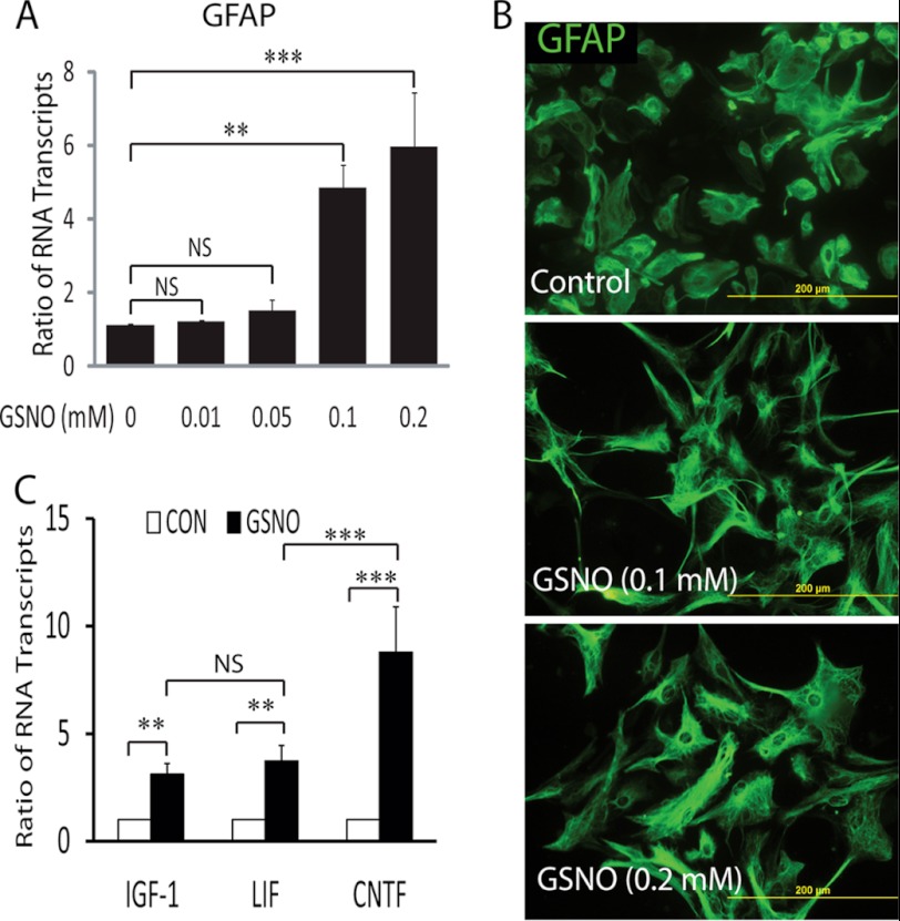
FIGURE 1.
Effect of GSNO treatment in astrocytes. Cortical astrocytes were plated (2000 × cm2) in 6-well cell culture plates or glass slide chambers. After 24 h, cells were treated with GSNO for another 24 h. A, the composite mean ± S.E. of four experiments depicts the ratio of GFAP to β-actin mRNA transcripts in treated astrocytes. B, representative fields of the slides (n = 5) depict the morphology of GFAP positive astrocytes determined by immunocytochemistry and fluorescence microscopy (magnifications ×400). C, the composite mean ± S.E. of three experiments depicts the ratio of IGF-1, LIF, and CNTF to β-actin mRNA transcripts in astrocytes treated with GSNO (0.2 mm) for 24 h. Statistical significance as indicated **, p < 0.01; ***, p < 0.001; NS, not significant.

