
FIGURE 1.
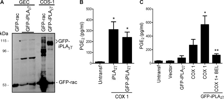
Expression and activity of M1 GFP-iPLA2γ WT. A, GECs were stably transfected, and COS-1 cells were transiently transfected with M1 GFP-iPLA2γ WT or a GFP-rac fusion protein for comparison. Lysates were immunoblotted with antibody to GFP. * denotes M1 GFP-iPLA2γ in GECs, which migrated slower than M1 GFP-iPLA2γ in COS-1 cells, possibly because of differential post-translational modifications in the two cell lines. B, COS-1 cells were transiently transfected with untagged full-length iPLA2γ, M1 GFP-iPLA2γ WT, and COX1. PGE2 production was measured in cell supernatants 24 h after transfection. Both GFP-tagged and untagged enzymes increased PGE2 release. *, p < 0.001 iPLA2γ + COX1 and GFP-iPLA2γ + COX1 versus untransfected (Untransf) cells, three experiments. C, COS-1 cells were untransfected or were transiently transfected with vector, M1 GFP-iPLA2γ WT, COX1, or M1 GFP-iPLA2γ WT + COX1. Expression of COX1 alone increased PGE2 release compared with control, whereas GFP-iPLA2γ + COX1 substantially amplified the increase in PGE2 production. This increase was inhibited by coincubation with 30 μm BEL (6 h). *, p < 0.001 GFP-iPLA2γ + COX1 versus untransfected or vector and p < 0.001 GFP-iPLA2γ + COX1 versus GFP-iPLA2γ, p < 0.05 GFP-iPLA2γ + COX1 versus COX1; **, p < 0.001 GFP-iPLA2γ + COX1 + BEL versus GFP-iPLA2γ + COX1, 7 experiments.