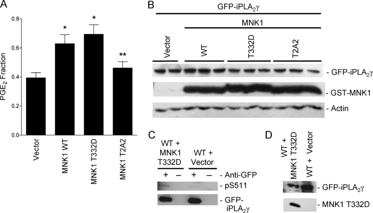
FIGURE 11.
Constitutively active MNK1 activates and phosphorylates iPLA2γ. COS-1 cells were transiently co-transfected with M1 GFP-iPLA2γ WT, COX1 and GST-MNK1 WT, GST-MNK1 T332D, and GST-MNK1 T2A2 or with empty vector. A, PGE2 release was measured 48 h after transfection. *, p < 0.05 MNK1 WT versus vector; *, p < 0.01 MNK1 T332D versus vector and **, p < 0.05 MNK1 T332D versus MNK1 T2A2, five experiments. In these experiments basal PGE2 release (vector + M1 GFP-iPLA2γ WT + COX1-transfected cells) was 164 pg/ml. B, lysates were immunoblotted with antibodies to GFP, GST, or actin. The blot shows comparable levels of expression. C, COS-1 cells were transiently co-transfected with M1 GFP-iPLA2γ WT and GST-MNK1 T332D or vector. After 48 h cells were treated with ionomycin (10 μm, 40 min) (ionomycin was included in these experiments to enhance the phosphorylation signal, although ionomycin did not independently induce phosphorylation; see Fig. 12). Cell lysates were immunoprecipitated with anti-GFP antibody (+) or nonimmune IgG in controls (−) and were immunoblotted with anti-(R/K)XX(pS/T) or anti-GFP antibodies. The blot shows enhanced phosphorylation of iPLA2γ Ser-511 (pS511) in MNK1 T332D transfected cells. D, total lysates of the above immunoprecipitation experiments blotted with anti-GFP or anti-GST (MNK1) antibodies are shown.

