
FIGURE 12.
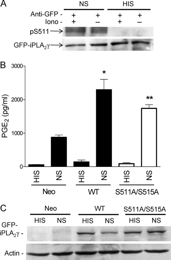
Complement induces phosphorylation of iPLA2γ on Ser-511. GECs were transiently transfected with M1 GFP-iPLA2γ WT. After 24 h cells were incubated with ionomycin (Iono, 5 μm) for 30 min (+) and then incubated with antibody and NS (3%, 40 min) or HIS in controls. A, cell lysates were immunoprecipitated with anti-GFP antibody (+) and were immunoblotted with anti-(R/K)XX(pS/T) or anti-GFP antibodies. The blot shows phosphorylation of Ser-511 (pS511) in NS-stimulated cells (with or without ionomycin). Phosphorylation is absent in the HIS-stimulated cells. B, GEC neo and GECs that express M1 GFP-iPLA2γ WT or GFP-iPLA2γ S511A/S515A (transient transfection) were incubated with antibody and complement as above. PGE2 production was amplified in NS-stimulated M1 GFP-iPLA2γ WT-expressing cells, whereas the amplification was smaller in GECs expressing the double mutant. *, p < 0.001 M1 GFP-iPLA2γ WT (NS) versus GEC-Neo (NS) and **, p < 0.05 M1 GFP-iPLA2γ WT (NS) versus GFP-iPLA2γ S511A/S515A (NS), three experiments. C, cell lysates were immunoblotted with antibodies to GFP or actin. The blot shows comparable levels of GFP-iPLA2γ expression.