FIGURE 4.

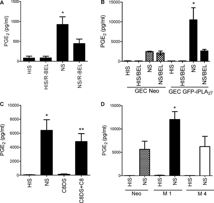
Complement induces production of PGE2 via iPLA2γ. A, shown is the role of endogenous iPLA2γ. Neo GECs were incubated with anti-GEC antiserum for 30 min at 22 °C in the presence or absence of the iPLA2γ-directed inhibitor R-BEL (10 μm). Cells were then incubated at 37 °C with 2% NS (to form C5b-9) or HIS in controls with or without R-BEL for 40 min. Then PGE2 production was measured in cell supernatants. Complement stimulated PGE2 production, and the increase was significantly attenuated by R-BEL. *, p < 0.0001 NS versus HIS and p < 0.001 NS versus NS/R-BEL, three experiments. B, complement-induced production of PGE2 is amplified in GECs that overexpress M1 GFP-iPLA2γ WT (GEC GFP-iPLA2γ). GECs that express M1 GFP-iPLA2γ WT were incubated with antibody and complement with or without BEL as above. M1 GFP-iPLA2γ WT markedly amplified complement-induced PGE2 production, and the increase was attenuated by BEL (30 μm). *, p < 0.001 GEC-GFP-iPLA2γ (NS) versus GEC-Neo (NS), p < 0.001 GEC-GFP-iPLA2γ (NS/BEL) versus GEC-GFP-iPLA2γ (NS), three experiments. C, PGE2 production is dependent on C5b-9 assembly. GECs that express M1 GFP-iPLA2γ WT were incubated with antibody and C8-deficient serum (C8DS) with or without purified C8. When C8DS was reconstituted with C8, PGE2 production amplified significantly. *, p < 0.0001 NS versus HIS; **, p < 0.001 C8DS+C8 versus C8DS, 3 experiments. D, M4 GFP-iPLA2γ is inactive in intact GECs. Neo GECs or GECs that stably express M1 GFP-iPLA2γ WT or M4 GFP-iPLA2γ were incubated with antibody and complement as above. *, p < 0.001 GEC-M1 GFP-iPLA2γ WT (NS) versus GEC-Neo (NS) and p < 0.001 GEC-M1 GFP-iPLA2γ WT (NS) versus GEC-M4 GFP-iPLA2γ (NS), three experiments.
