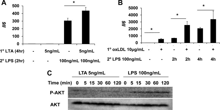
FIGURE 7.
Cross-priming of murine macrophages by lipoteichoic acid and oxLDL. A, WT BMDM were treated with medium control, 5 ng/ml LTA, 100 ng/ml LPS, or an initial LTA (5 ng/ml) for 4 h, followed by LPS (100 ng/ml) for 2 h. The levels of expressed Il-6 were measured by real-time RT-PCR. B, WT BMDM were treated with medium control, 10 μg/ml oxLDL, 100 ng/ml LPS for 4 h, or an initial oxLDL (10 μg/ml) for 4 h, followed by LPS (100 ng/ml) for 4 h. The levels of expressed Il-6 were measured by real-time RT-PCR. C, WT BMDM were treated as indicated in the legend to Fig. 7. The levels of phosphorylated Akt (p-AKT) and total Akt were detected by Western blot. *, p < 0.05.

