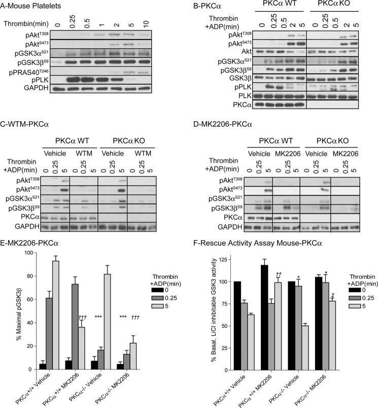
FIGURE 3.
Thrombin-stimulated GSK3 phosphorylation and inhibition of GSK3α/β activity is delayed in platelets from PKCα KO mice. Washed PKCα+/+ (PKCα WT, A–F) and PKCα−/− (PKCα KO, B–F) mouse platelets were stimulated with 0.2 unit/ml thrombin (A) or 0.2 unit/ml thrombin+10 μm ADP (B–F) for the indicated times. Platelets were extracted in NuPAGE sample buffer followed by immunoblotting with the indicated antibodies (A–D). Results (A–D) are representative of at least three independent experiments. The bar graph (E) depicts densitometric analysis of GSK3β phosphorylation of the data shown in D. Data (mean ± S.E. (error bars), n = 5) are expressed as the percentage of maximal GSK3 phosphorylation induced by thrombin+ADP in PKCα WT mouse platelets. Star notation (E: ***, p < 0.001) indicates a significant difference compared with 0.25-min thrombin+ADP treatment in vehicle-treated PKCα WT platelets whereas dagger notation (E: †††, p < 0.001) indicates a significant difference compared with 5-min thrombin+ADP treatment in vehicle-treated PKCα WT platelets. Alternatively, platelets were extracted in Nonidet P-40 (F) followed by analysis of in vitro GSK3 activity using the substrate peptide RRAAEELDSRAGS(P)PQL. In vitro kinase activity is expressed as a percentage of GSK3 activity obtained in the absence of thrombin and ADP (mean ± S.E., n = 5). Star notation (F: *, p < 0.05) indicates a significant difference compared with 0.25-min thrombin+ADP treatment in vehicle-treated PKCα WT platelets; dagger notation (††, p < 0.01) indicates a significant difference compared with 5-min thrombin+ADP treatment in vehicle-treated PKCα WT platelets, whereas double dagger notation (‡, p < 0.01) indicates a significant difference compared with 5-min thrombin+ADP treatment in vehicle-treated PKCα KO platelets.

