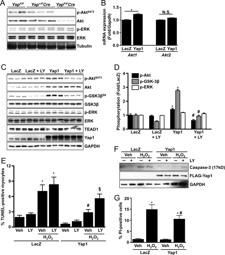
FIGURE 3.
Yap1 promotes cardiomyocyte survival through up-regulation of Akt. A, representative immunoblots detect p-Akt(S473), total Akt, p-ERK(T202/Y204), and total ERK from Yap+/F, Yap+/FCre, and YapF/FCre hearts. B, neonatal rat cardiomyocytes were transduced with LacZ control or Yap1 adenovirus, and quantitative real-time-PCR was performed to determine mRNA expression of Akt1 and Akt2. Results are normalized to Gapdh expression. *, p < 0.05. N.S. = not significant. C, cardiomyocytes were transduced with LacZ or Yap1 adenovirus and treated with LY294002 (10 μm) or vehicle. Representative immunoblots are shown. D, quantification of results was obtained from C. *, p < 0.05 versus LacZ. #, p < 0.05 versus Yap1. E, cardiomyocytes were transduced with LacZ or Yap1 adenovirus and treated with LY294002 (10 μm) or vehicle. Cells were then treated with vehicle or H2O2 (100 μmol/liter), and apoptosis was evaluated by TUNEL. Values are the means ± S.E. *, p < 0.05 versus LacZ + vehicle. #, p < 0.05 versus LacZ + H2O2 + vehicle. §, p < 0.05 versus Yap1 + H2O2 + vehicle. F, cardiomyocyte apoptosis was also determined by detection of cleaved caspase-3 (17kD). A representative immunoblot is shown. G, cardiomyocyte viability was determined by propidium iodide (PI) uptake after H2O2 (100 μmol/liter) treatment. Values are the means ± S.E. *, p < 0.05 versus LacZ + vehicle. #, p < 0.05 versus LacZ + H2O2.

