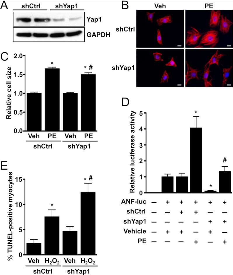
FIGURE 5.
Depletion of Yap1 attenuates cardiomyocyte hypertrophy and promotes apoptosis. A, shown is a representative immunoblot demonstrating endogenous Yap1 knockdown using adenoviral shRNA. Cardiomyocytes were infected with scrambled control (shCtrl) or Yap1-targeted (shYap1) virus, and cells were harvested 72 h later. B, cardiomyocytes were transduced with shCtrl or shYap1, treated with phenylephrine (PE; 100 μm) or vehicle, and stained to detect troponin T. Scale bars, 10 μm. C, quantification of the cardiomyocyte surface area is shown. Values are the means ± S.E. *, p < 0.05 versus shCtrl + vehicle. #, p < 0.05 versus shCtrl + phenylephrine. D, cardiomyocytes were transfected with shCtrl or shYap1 in combination with ANF-luc reporter plasmid. Cells were treated with phenylephrine (100 μm) or vehicle control, and luciferase activity was determined. Values are the means ± S.E. *, p < 0.05 versus shCtrl + vehicle. #, p < 0.05 versus shCtrl + phenylephrine. E, cardiomyocytes were transduced with shCtrl or shYap1 adenovirus and treated with vehicle or H2O2 (100 μmol/liter), and apoptosis was evaluated by TUNEL. Values are the means ± S.E. *, p < 0.05 versus shCtrl + vehicle. #, p < 0.05 versus shCtrl + H2O2.

