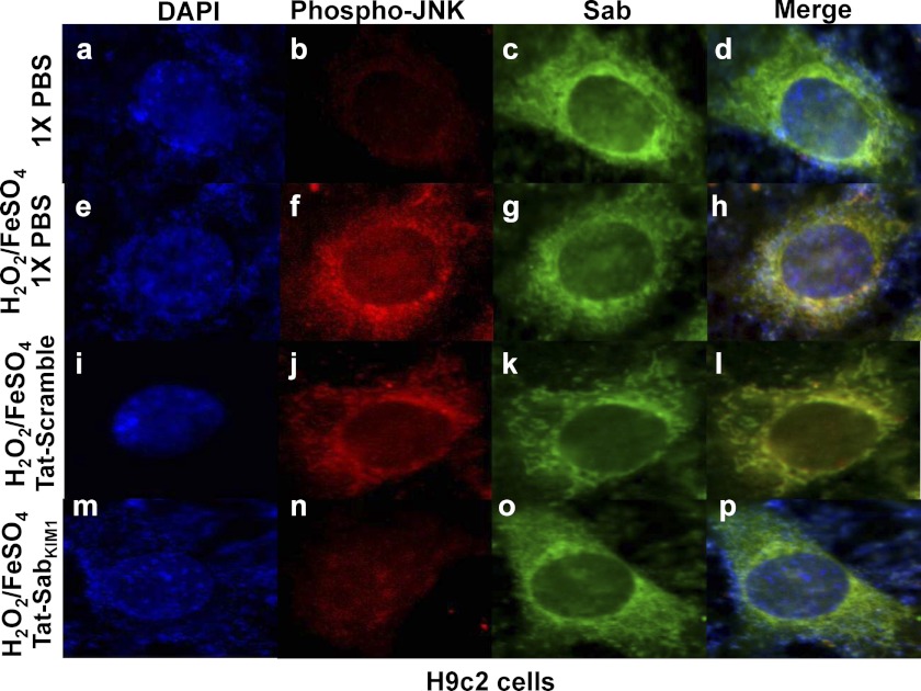
FIGURE 3.
Immunofluorescence of phospho-JNK and Sab in H9c2 cells. Immunofluorescence of H9c2 cells demonstrates JNK co-localization with mitochondrial Sab in the presence of H2O2/FeSO4. Unstressed cells treated with 1× PBS and cells treated with 100 μm H2O2/FeSO4 for 20 min in the presence and absence of Tat-scramble or Tat-SabKIM1 were incubated with antibodies specific for active JNK (Phospho-JNK) and Sab. Cells were then incubated with fluorescently conjugated secondary antibodies as described under “Experimental Procedures.” Cells were stained with DAPI following antibody incubations. Images were acquired using fluorescence microscopy at ×100 magnification.

